
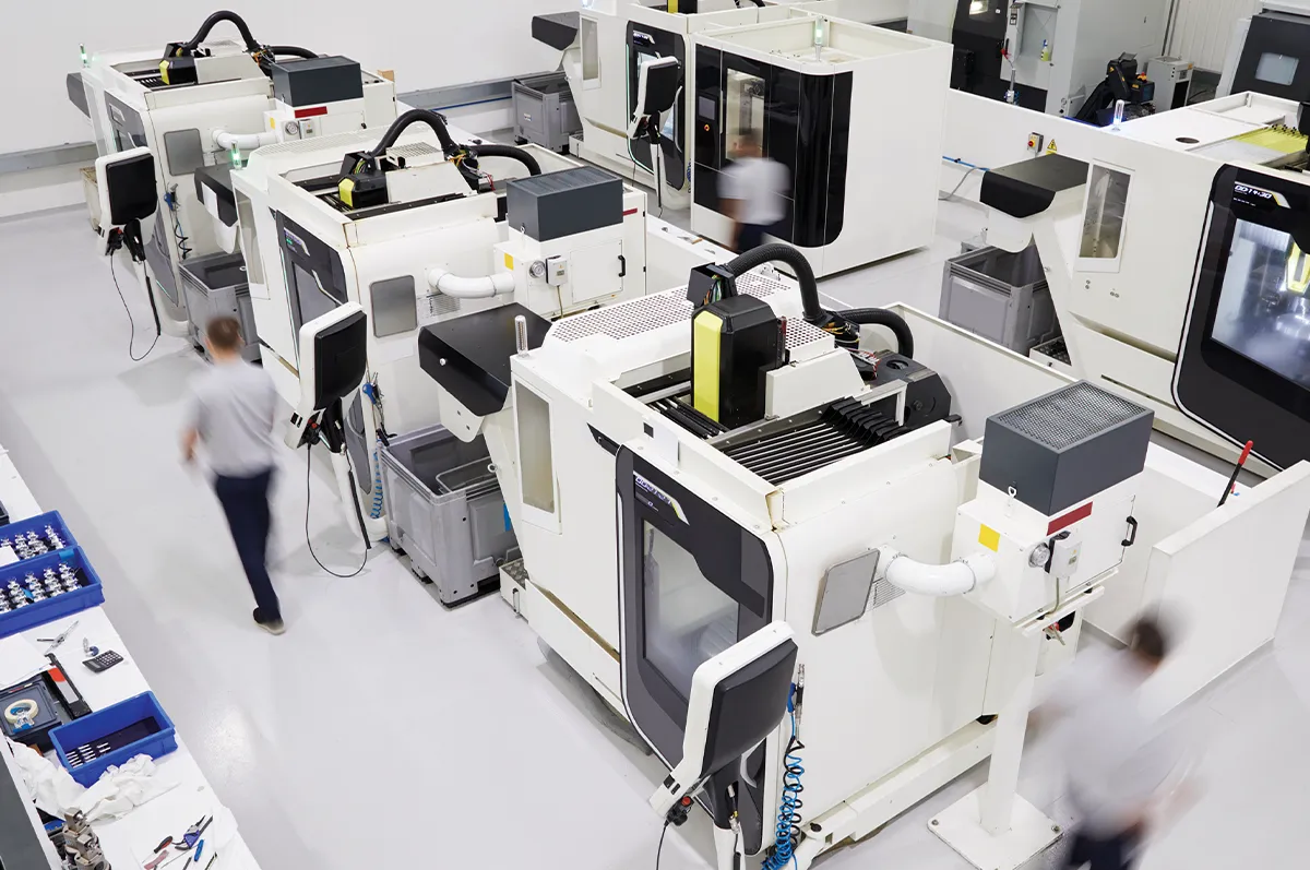
Next-Gen Implant Manufacturing: Integrating Digital Workflows and Patient-Specific Customization
The medical device industry is undergoing a structural paradigm shift. Traditional “one-size-fits-all” orthopedic and dental implants are rapidly giving way to patient-specific implants (PSIs). Driven by high-resolution diagnostic imaging, artificial intelligence (AI), and additive manufacturing (3D printing), the modern digital workflow enables medical device manufacturers, surgeons, and healthcare facilities to design and fabricate implants tailored precisely to an individual’s unique anatomical geometry.
This digital ecosystem minimizes operating room time, lowers post-operative complication rates, and accelerates patient recovery. Understanding how to integrate digital workflows with patient-specific customization is becoming essential for medical device engineering, surgical planning, and orthopedic supply chains worldwide.
High-Precision Diagnostic Imaging and Data Acquisition
The patient-specific manufacturing workflow begins at the clinic with diagnostic radiology. Standardized medical imaging modalities convert a patient’s physical anatomy into a machine-readable digital dataset.
- Computed Tomography (CT) & CBCT: High-resolution CT scans serve as the gold standard for hard-tissue geometry, capturing bone density and structural boundaries with sub-millimeter precision. Cone Beam CT (CBCT) is widely deployed for craniomaxillofacial (CMF) and dental reconstructions.
- Magnetic Resonance Imaging (MRI): Used alongside CT data when soft-tissue boundaries, such as cartilage, ligaments, or nerve pathways, influence implant placement.
- Intraoral & Optical Surface Scanning: Provides high-fidelity surface topography for dental restorations and surface-level maxillofacial mapping.
Data sets are stored and transmitted via DICOM (Digital Imaging and Communications in Medicine) protocol standards, establishing an interoperable foundation for downstream engineering software
Image Segmentation and AI-Assisted Anatomical Reconstruction
Once DICOM files enter the engineering workflow, they undergo image segmentation—a process that isolates targeted anatomical structures from surrounding tissues.
+——————-+ +——————-+ +——————-+
| DICOM Acquisition| —> | AI Segmentation | —> | 3D Mesh / CAD |
| (CT / MRI / CBCT) | | (Threshold/Deep L)| —> | (STL / OBJ / 3MF) |
+——————-+ +——————-+ +——————-+
Digital Processing Steps
- Hounsfield Unit (HU) Thresholding: Assigns density values to bone tissue to separate hard structures from soft tissue and background artifacts.
- Machine Learning Automation: AI and deep-learning segmentation algorithms automatically detect anatomical landmarks, identify pathology, and perform volumetric segmentation in minutes—a process that historically took several hours of manual tracing.
- Surface Mesh Generation: Cleaned anatomical data is converted into 3D triangular surface meshes, standardizing the format as STL, OBJ, or 3MF files for CAD software compatibility.
Surgical Design, Generative Optimization, and VSP
Patient-specific implant design combines biomedical engineering with surgeon-led Virtual Surgical Planning (VSP). Rather than altering host bone to fit off-the-shelf implant geometry, the implant is engineered to match the patient’s existing anatomical contours.
| Parameter | Standard Mass-Produced Implants | Patient-Specific Custom Implants |
| Fit & Alignment | Requires intraoperative bone reshaping | Form-fitted to patient-specific CAD geometry |
| Porosity & Lattice | Uniform coating or solid metal | Tailored porous lattice structure via 3D printing |
| Surgical Efficiency | Higher trial-and-error adjustment time | Streamlined using pre-designed surgical guides |
| Bone Ingrowth (Osseointegration) | Dependent on mechanical press-fit | Optimized pore size (300–600 µm) and interconnectivity |
Generative Design & Lattice Structures
Advanced CAD tools utilize generative algorithms and topology optimization to create complex internal lattice structures (e.g., Gyroid or Diamond TPMS lattices). These porous configurations offer two distinct advantages:
- Biomechanical Stiffness Matching: Reduces the modulus of elasticity of solid metals (such as Titanium) down to match natural trabecular bone (~0.5 to 3.0 GPa), significantly mitigating the risk of stress shielding and subsequent bone resorption.
- Osseointegration: Interconnected pore channels ranging between 300 and 600 micrometers encourage vascularization and long-term biological fixation.
Additive Manufacturing and Material Selection
Additive manufacturing (AM) serves as the primary production mechanism for custom medical implants, enabling the fabrication of complex geometric shapes impossible to manufacture using traditional multi-axis CNC milling.
Metal Additive Technologies
- Selective Laser Melting (SLM) / Laser Powder Bed Fusion (L-PBF): Uses high-power fiber lasers to melt microscopic metal powders layer-by-layer.
- Electron Beam Melting (EBM): Utilizes an electron beam in a high-vacuum environment, reducing residual stresses and processing reactive metals at elevated thermal states.
Biocompatible Materials
- Ti-6Al-4V ELI (Grade 23 Titanium): The industry benchmark for load-bearing orthopedic, cranial, and dental implants due to its high strength-to-weight ratio, fatigue resistance, and biocompatibility.
- PEEK (Polyetheretherketone) & PEKK: Radiolucent thermoplastic polymers utilized in cranial reconstruction and spinal cages due to their closeness in elasticity to cortical bone.
- Tantalum: Highly corrosion-resistant metal known for its biological response and high friction coefficient in primary joint revisions.
Post-Processing, Quality Assurance, and Regulatory Compliance
Additive manufacturing is only one phase of the overall implant fabrication life cycle. Medical devices undergo strict post-processing and quality assurance steps to ensure mechanical performance and biological safety.
- Stress Relief & Heat Treatment: Implants undergo Hot Isostatic Pressing (HIP) or thermal annealing to relieve internal thermal stresses, close micro-voids, and increase fatigue life.
- Powder Removal & Surface Finishing: Trapped unfused powder is purged via ultrasonic cleaning and automated air blowing. Surface etching or electropolishing refines surface roughness to optimize cellular attachment.
- Validation & Metrology: Industrial Computed Tomography (Micro-CT) and coordinate measuring machines (CMM) inspect the finished implant to verify dimensional tolerances against original patient CAD models.
- Sterilization & Packaging: Implants undergo validation in ISO Class 7 or 8 cleanroom environments, followed by Gamma Irradiation, Ethylene Oxide (EtO), or Autoclave sterilization prior to surgical delivery.
Key Benefits for Modern Healthcare Systems
Integrating digital workflows with customized manufacturing provides structural efficiency across the clinical continuum:
- Reduced Intraoperative Risk: Pre-planned virtual cuts and custom surgical cutting guides reduce time spent under general anesthesia.
- Superior Anatomical Fit: Custom-contoured implants eliminate the need for manual intraoperative plate bending or aggressive bone reaming.
- Improved Long-Term Outcomes: Custom pore architectures improve primary biological fixation, reducing revision surgery rates in complex oncological, trauma, and orthopedic cases.
As machine learning algorithms, intraoperative navigation, and additive manufacturing processes continue to converge, end-to-end digital implant manufacturing stands at the forefront of personalized, value-based healthcare.