
The Evolution of Implantology: How Advanced Surface Technology and Biocompatibility Drive Osseointegration
The field of implantology has experienced a profound shift over the past few decades. Modern dental and orthopedic implants are no longer passive mechanical anchors inserted into bone; they are highly engineered, bio-interactive devices designed to integrate seamlessly with human tissue. At the center of this transformation lies osseointegration—the direct structural and functional connection between living bone and the surface of a load-bearing implant.
Achieving rapid, predictable, and long-lasting osseointegration depends heavily on two critical factors: advanced surface technology and material biocompatibility. As clinical demands push toward shorter healing times and immediate loading protocols, implant manufacturers are continuously innovating at the microscopic and nanoscopic levels.
Understanding Osseointegration: The Biological Foundation
First introduced by Professor Per-Ingvar Brånemark in the 1960s, osseointegration revolutionized restorative dentistry and orthopedics. When an implant is placed into the host bone, a complex sequence of biological events is initiated:
- Hemostasis and Blood Contact: Within seconds, blood covers the implant surface, forming a fibrin matrix.
- Inflammatory Response: Cytokines and growth factors are released, recruiting osteoprogenitor cells to the site.
- Bone Formation (Osteogenesis): New bone matrix is synthesized on the implant surface (contact osteogenesis) and from the surrounding bone wall (distance osteogenesis).
- Remodeling: Over months, woven bone transforms into mature, lamellar bone capable of withstanding functional loads.
The speed and quality of this process depend directly on how osteoblasts (bone-forming cells) interact with the implant’s outermost layer.
The Role of Biocompatibility: Biomaterials in Modern Implantology
Biocompatibility is the fundamental requirement for any implant material. It defines the ability of a material to perform with an appropriate host response in a specific application.
Titanium and Its Alloys
Grade 4 commercially pure titanium (cpTi) and Ti-6Al-4V ELI (Extra Low Interstitial) alloy remain the gold standard in implant manufacturing. Titanium owes its exceptional biocompatibility to its spontaneous formation of a passive titanium dioxide ($\text{TiO}_2$) oxide layer (typically 2 to 10 nm thick) when exposed to oxygen. This oxide layer prevents corrosion, resists chemical degradation, and provides a favorable surface for cell attachment.
Ceramic Alternatives: Zirconia
In response to increasing demands for metal-free solutions and enhanced esthetics, Yttria-stabilized Tetragonal Zirconia Polycrystals (Y-TZP) have emerged as a viable alternative. Zirconia offers high flexural strength, soft-tissue friendliness, low plaque accumulation, and excellent tooth-like esthetics, making it popular in the anterior zone.
Surface Technology: Engineering the Interface
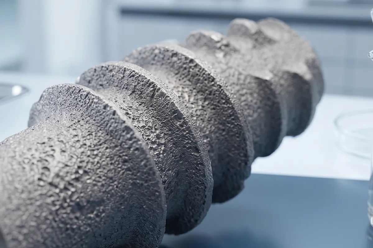
While material selection establishes biocompatibility, surface modification dictates the speed and strength of osseointegration. Surface topography is categorized into three physical scales: macro, micro, and nano.
Micro-Roughness and Mechanical Interlocking
Pioneering implants utilized smooth, machined surfaces. Methods to achieve this include:
- Sandblasting and Acid-Etching (SLA): Large-grit sandblasting creates macro-pits, followed by acid-etching to overlay micro-pits.
- Resorbable Blast Media (RBM): Uses biocompatible materials like calcium phosphate instead of alumina, leaving a clean, textured surface.
Micro-roughness increases the total surface area available for bone-to-implant contact (BIC) and provides mechanical anchorage for early stability.
Nanotechnology and Cellular Behavior
Nanotopography operates at the scale of proteins and cell receptors. Nano-engineered features—such as titanium dioxide nanotubes—mimic the natural extracellular matrix, promoting osteoblast adhesion, cell differentiation, and gene expression dedicated to matrix mineralization.
Hydrophilicity: The New Frontier in Early Healing
Historically, titanium surfaces exposed to ambient air absorbed hydrocarbons, rendering them hydrophobic (water-repelling). Modern surface technology counteracts this through hydrophilic (superhydrophilic) modification.
Hydrophilic surfaces feature an extremely low contact angle with liquids ($< 10^\circ$). When exposed to blood during surgical placement:
- Blood flows instantly into micro- and nano-textured crevices.
- Protein adsorption (such as fibronectin and vitronectin) is accelerated.
- Clot formation is stabilized directly against the surface.
By enhancing early biological activity, hydrophilic surfaces significantly shorten the critical healing period (from 6–8 weeks down to 3–4 weeks), reducing failure rates during the vulnerable “dip” in mechanical stability when primary stability transitions to secondary stability.
Comparative Summary: Implant Surface Modifications
| Surface Type | Processing Method | Primary Biological Advantage | Typical Healing Window |
| Machined / Smooth | Lathe turning | Minimal friction during insertion | 3–6 Months |
| Micro-Rough (SLA/RBM) | Sandblasting & Acid Etching | Increased mechanical keying and higher BIC | 6–8 Weeks |
| Hydrophilic Micro-Rough | Nitrogen-protected storage or plasma cleaning | Rapid clot stabilization & early osteogenesis | 3–4 Weeks |
| Nano-Structured (Nanotubes) | Anodization / Nano-coating | Cellular guidance & anti-bacterial properties | Under Investigation / Rapid |
Future Trends: Smart Surfaces and Antimicrobial Coatings
As implantology advances, the focus is shifting from simple bone integration to preventative and active healing surfaces.
- Antimicrobial and Anti-Infective Surfaces: Peri-implantitis remains a leading cause of long-term failure. Innovations include silver nanoparticle coatings, bio-inspired anti-fouling topographies, and localized antibiotic delivery systems embedded in porous oxides.
- Growth Factor Immobilization: Coating implant surfaces with Bone Morphogenetic Proteins (BMPs) or specific peptide sequences (like RGD peptides) directly stimulates cell recruitment without waiting for endogenous signal cascades.
- Additive Manufacturing (3D Printing): Selective Laser Melting (SLM) allows the creation of porous titanium structures that match the elastic modulus of trabecular bone, minimizing stress shielding while encouraging bone endogrowth.